The Ellison Institute addresses pressing challenges in cancer care and promotes innovation through the integration of clinically driven science and applied engineering. Guided by our Chief Science and Innovation Officer, Dr. Jerry S.H. Lee, the Ellison Institute’s overarching objective is the rigorous and rapid deployment of novel technologies to enhance clinical practice, diagnostics, and research. The combination of our public-private partnerships, our bi-directional feedback between our clinic and research enterprises, and our robust data from an ever-growing biorepository enables the Ellison Institute to accelerate advancements in research and patient care. As a biomedical and technology hub, we collaborate with leading physicians, scientists and companies to propel vital cancer research initiatives along the analytical pipeline to create clinically impactful interventions. Expert scientists, physicians, and thought leaders from around the world are invited to become visiting faculty at the Institute and collaborate with our multidisciplinary resident scientists. By uniting international experts from diverse disciplines under one roof, the Ellison Institute aims to deliver insight and ingenuity to surmount hurdles that have been plaguing researchers and physicians for years.
Leveraging Collaboration and Interdisciplinary Science
"I believe we can advance our research to the next level, allowing the most effective treatments to benefit patients who are in urgent need of new therapies today."
–– David B. Agus, MD
MULTISCALE BIOLOGY LAB
DRUG DISCOVERY LAB
MOLECULAR ANALYTICS LAB
APPLIED THERAPEUTICS LAB
MICRo - ENVIRONMENT LAB
biomimetic models lab
proving ground
immersive visualization lab
integrative microscopy lab
ARTIFICIAL INTELLIGENCE IN MEDICINE
The Artificial Intelligence in Medicine Lab utilizes machine learning to unlock new clues in existing datasets and draw unprecedented insights to enhance cancer diagnostics and treatment.
LEFT IMAGE: A breast cancer pathology slide;
RIGHT IMAGE: The same slide overlaid with an AI-generated “heat map” identifying zones of clinical importance. (Photo credit: Rishi Rawat, PhD)
Accurate cancer diagnoses that lead to timely, effective treatments are dependent upon high-confidence pathology reports. However, traditional pathology’s utility is limited by variation in tissue processing across laboratories, limitations in human perception, and simple differences of opinion. Moreover, the molecular staining techniques required for precision medicine are expensive and challenging to execute, resulting in considerable sources of variability in the cancer diagnosis process. Our researchers at the Ellison Institute are leveraging the power of machine learning algorithms to streamline and standardize cancer diagnostics and, ultimately, to remove human error from the equation.
Ellison Institute scientists have already made significant headway using artificial intelligence to enhance breast cancer diagnostics by predicting which patients will likely respond to a hormonal therapy targeting the estrogen receptor pathway. The architecture of breast cancer that contains cells expressing the estrogen receptor varies minutely from that containing cells lacking the receptor; although the human eye cannot detect these differences, a computer can. Our scientists have demonstrated that their deep learning algorithm can analyze pathology images of breast cancer cells and predict estrogen receptor status based on tissue architecture alone, without requiring the usual time-consuming staining that reveals estrogen receptors to the human eye. These groundbreaking results hold immense promise to complement the role of a human pathologist, reduce diagnostic errors and ultimately help deliver results to patients with unprecedented accuracy and efficiency.
In addition to employing artificial intelligence to enhance patient care, our researchers have devoted their energies to advancing the way data scientists around the world can train digital pathology algorithms to achieve better outcomes. Deep learning algorithms require extensive training on large datasets before they can churn out predictions with consistency and high confidence; Ellison Institute researchers have devised a technique called “tissue fingerprinting” to significantly reduce the size of training datasets while increasing predictive accuracy. Our institute is working to continually hone these tools to revolutionize diagnosis and treatment planning while simultaneously serving as a model for digital pathology researchers around the world.
MULTISCALE BIOLOGY LAB
The Multiscale Biology Lab combines biological, physical and mathematical approaches to surpass traditional cancer research capabilities and study tumor growth, metastasis and drug resistance from the subcellular level all the way to the full-body scale.
IMAGE: This is an image of a colorectal cancer organoid derived from patient tissues. The nuclei are stained using DAPI (blue), proliferating cells are depicted with Ki67 staining (pink), and actin filaments are stained with phalloidin (green). (Photo credit: Seungil Kim, PhD)
Much of traditional cancer biology focuses on studying the growth and proliferation of small, homogeneous populations of tumor cells cultured within the confines of a two-dimensional Petri dish. While this type of research is essential to the field of oncology, when used by itself to study cancer it offers a limited scope of understanding and fails to answer a variety of essential questions. How do cancer cells interact with the healthy, supportive cells that surround them in three dimensions? How does the tumor microenvironment change when drugs are introduced? How do the inherent biophysical characteristics of a tumor and its microenvironment contribute to or impede metastasis? To answer these key questions, we have to study cancer across a variety of scales and involve other disciplines such as math and physics to more accurately model cancer’s behavior at the molecular, cellular, tumor, organ and systemic levels.
Traditional biology meets mathematical modeling
One of our mathematical oncology initiatives at the Ellison Institute combines biological and computational approaches to dynamically model colorectal cancer growth, track microenvironmental interactions and evaluate candidate therapies. To account for the subcellular processes driving cancer growth and drug response, EI Member Stacey Finley, PhD pulls together experimental data reflecting biochemical reactions, molecular signaling and cellular metabolism within both cancer cells and neighboring healthy cells found in the tumor microenvironment. Finley synthesizes this information to create algorithms that model cellular behavior. Our collaborator Paul Macklin, PhD at Indiana University uses Finley’s data to inform his own algorithms that model entire tumors and account for microenvironmental factors like oxygen levels, nutrient availability and cell-cell signaling among cancer cells and between healthy and malignant cells. Finally, EI faculty member Shannon Mumenthaler, PhD grows three-dimensional cell culture models called organoids, which contain both cancerous and non-cancerous surrounding cells, to create biological analogs for the team’s computational models. Taken together, these multiscale models create a much more realistic surrogate for in vivo tumor growth than traditional two-dimensional cell cultures, and their parameters can be modified to test hypotheses with unprecedented capabilities.
One of the most exciting applications of this approach is the rapid, inexpensive simulation of thousands of drug combinations to identify novel therapeutic approaches against colorectal cancer. It simply would not be feasible to create biological models to test every possible drug regimen in an outright fashion, but computational models can evaluate multitudes of drug combinations and doses to select a small group of candidates for further testing. Once we develop and test the top predictions for their efficacy in vitro using our organoid models, we can feed those outcomes back into our algorithms to enhance their predictive capabilities and generate new scientific hypotheses.
Beyond testing existing drugs, we ultimately hope to use our models to develop new therapeutics targeting the microenvironment––a relatively novel concept for the field of oncology, which has historically focused its drug development efforts on the malignant cells themselves. Using our mathematical models, we can test various perturbations of nutrient levels to identify microenvironmental changes that most significantly impact tumor survival. Our goal is to both identify clinically useful biomarkers to predict drug response and screen targeted therapies that fight cancer by disrupting key aspects of the microenvironment. Our researchers have already made discoveries highlighting the important role of microenvironmental factors in tumor evolution and drug resistance, and we hope that this work will continue to draw attention to the often-overlooked importance of the microenvironment in understanding and fighting cancer.
The physics of metastasis
Much of cancer’s behavior cannot be explained by biology alone. Metastasis, for example, has long perplexed cancer biologists working to understand which cells hold metastatic potential, which are likely to survive their journeys out of their primary tumors and which select few will then be able to colonize and form new tumors in distant locations. To better address the questions of metastasis that have stumped scientists for decades, the Ellison Institute is borrowing brainpower from the fields of engineering and physics to approach these conundrums from a different angle and ultimately accelerate scientific breakthroughs.
One very important yet minimally understood biophysical property of cancer cells is their viscoelasticity, which describes the way cells respond when physical forces act upon them. How resistant are they to the pressure changes and shear forces they must undergo in order to leave their tissue of origin, enter the bloodstream and ultimately engraft in other tissues? When a cancer cell leaves its relatively stable, low-force primary tumor and enters the rapidly moving bloodstream, it experiences shear forces at that solid-liquid interface that are equivalent to the force a six-foot adult male would experience while trying to catch a 50-pound bag of sand with one hand. How does a cancer cell avoid getting ripped to shreds during that process? If it does manage to stay intact, what allows it to engraft and initiate a new tumor in an organ with distinctly different biophysical properties, such as when breast cancer metastasizes to the bones, liver, lungs, or brain?
Ellison Institute Chief Science and Innovation Officer Jerry S.H. Lee, PhD, a pioneer in the field of cancer biophysics, is currently leading a partnership with the biotechnology company Travera and the United States Department of Defense to study the nuances of cancer’s biophysical properties and find answers to these perplexing questions. Historically, one of the biggest obstacles when studying the biophysical properties of cancer has been procuring fresh, high-quality tissue samples to analyze; the Department of Defense’s APOLLO Biophysics Information Transfer study is transforming the field by procuring fresh surgical samples specifically prepared for researchers like us to use when studying the biophysical and biochemical properties of tumors. Using high-quality tissue samples from the Department of Defense, our scientists can employ Travera’s innovative technology to measure qualities such as viscoelasticity, pliability and resistance to force changes. With this information, we can identify the tools cancer uses to spread throughout the body and put a stop to that process.
In addition to our biophysics partnership with Travera and the Department of Defense, EI scientists are currently collaborating with researchers at the USC Viterbi School of Engineering to explore ways to selectively kill cancer cells based on the fact that they are “softer” and more pliable than their healthy counterparts. Their approach uses highly focused ultrasound waves to destroy malignancies while sparing the surrounding tissue, and the hope is that this technique will one day be used to both eradicate primary tumors and ablate metastatic lesions when they arise.
DRUG DISCOVERY LAB
The Drug Discovery Lab combines approaches from scientific disciplines such as chemistry, biology, physics and mathematics to accelerate drug development efforts, optimize cancer treatment and improve patient outcomes.
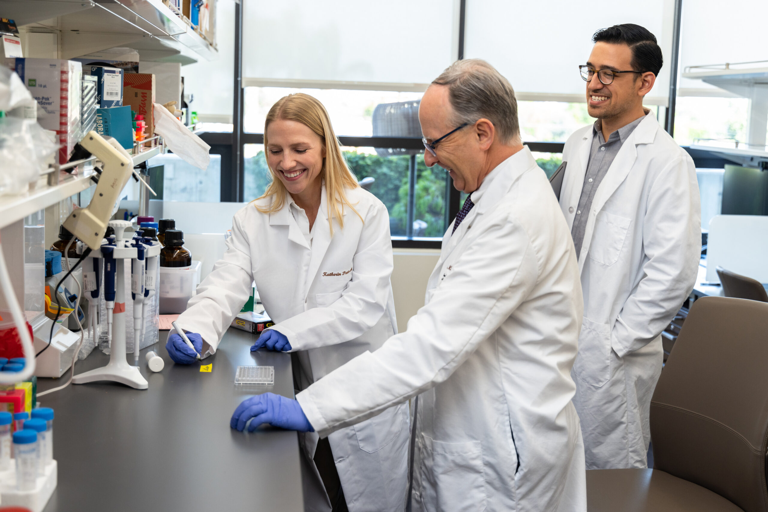
IMAGE: Ellison Institute Drug Discovery Lab members Katherin Patsch, PhD, and Seungil Kim, PhD, analyze patient-derived tumor samples in response to potential drug candidates using confocal microscopy.
To strengthen the translational nature of our research at the Ellison Institute, we have established the Drug Discovery Lab to develop novel therapeutics that achieve robust and long-lasting anti-cancer activity while minimizing unwanted side effects.
One of our most groundbreaking projects focuses on the development of an anti-androgen drug to treat castrate-resistant prostate cancer. Prostate cancer’s growth is driven by the androgen receptor (AR) pathway, which is activated by the male sex hormone testosterone. In what is known as castrate-resistant prostate cancer, disease progresses even after testosterone signaling has been blocked through prostate removal surgery or drug therapy. Most of these cancers still rely on the AR pathway to grow, meaning they have evolved ways to activate the pathway even in the absence of testosterone. These patients usually respond to recently developed antiandrogen drugs that directly target the AR. However, some patients must discontinue therapy due to side effects, and virtually all patients who continue therapy develop resistance within months. Thus, there is an urgent need to develop new antiandrogens to extend patients’ lives.
Ellison Institute scientists in the Drug Discovery Lab have partnered with EI Member Charles McKenna, PhD, director of the USC Center for Drug Discovery, to synthesize novel drugs that inhibit AR. Using advanced imaging technologies from Olympus and fluorescently labeled cancer cells developed at EI, we are able to screen hundreds of drug candidates in a day and visualize each compound’s activity on a subcellular level. Olympus’s machinery allows us to visualize in great detail where each drug intercepts the AR pathway, thereby enabling us to not only observe which drugs kill prostate cancer cells, but to also understand why those particular drugs are effective. Our scientists can in turn use that knowledge to identify new modes of drug action against the AR pathway to ultimately improve therapeutic outcomes.
Our team recently made the exciting discovery that a particular class of antiandrogens can act as either inhibitors or activators of the AR pathway––a finding that flies in the face of accepted knowledge of AR signaling in prostate cancer.
As our team moves forward with drug development, we will use our advanced imaging capabilities in conjunction with this new drug series to lead the scientific community toward a deeper understanding of AR pathway regulation. We hope these efforts will lead to new therapeutics that provide durable responses against castrate-resistant prostate cancer.
MOLECULAR ANALYTICS LAB
The Molecular Analytics Lab combines biological, chemical and physical analysis techniques to characterize tissue samples, monitor patients’ therapeutic responses and provide comprehensive snapshots of both healthy and diseased states.
IMAGE: Ellison Institute Director of Technical Operations Jonathan Katz, PhD, performs maintenance on the mass spectrometry instrumentation.
Cancer is often considered a genetic disease, but genomic data alone paints a very limited picture of cancer’s complexity, the variability between patients and the dynamic changes across different disease timepoints. DNA serves as a cellular blueprint, but additional layers of information such as gene activation, protein expression levels, molecular signaling and metabolic activity are what breathe life into the picture and enable us to define states of disease and wellbeing for our patients.
The Molecular Analytics Lab seeks to identify biomarkers and metabolic signatures in blood and tissue samples that clinicians can use to plan treatment approaches, measure patients’ responses to drugs and monitor health and disease progression over time. Our scientists can in turn use these metabolic profiles to track the way tumors evolve and respond to various types of therapies. Our goal is to provide patients with comprehensive genomic, proteomic and metabolomic analyses to thoroughly characterize their diseases and give our clinicians the best possible information to make life-saving decisions.
Currently, our team is employing sophisticated instrumentation from Agilent Technologies to perform robust molecular analyses on various types of blood cells to get a snapshot of what those circulating cells have encountered on their journeys throughout the body. We then extrapolate information from a series of metabolic snapshots to paint a more vivid “panoramic picture” that tells us unprecedented details about a patient’s overall health status. Other Ellison Institute research involves analyzing blood samples immediately after administering chemotherapy to determine quantitatively and quickly whether a patient is responding to a drug regimen.
Scaling molecular analytics workflows for use across the oncology landscape requires accounting for every preanalytical variable that could affect data outputs. Metabolic states change rapidly, and there is a brief window of time between when tissue is collected from the body and when samples start to degrade. Moreover, individual labs’ nuances in the way they handle and process samples before running tests can fundamentally skew results and render it difficult, if not impossible, to compare data insights across labs. Rigorous data collection standards are therefore essential to ensuring the information pulled from molecular analysis is accurate, consistent and clinically meaningful in a variety of practice settings.
The Ellison Institute has set out to help refine data collection standards in biomedicine through a partnership with the United States Department of Defense. Ellison Institute scientists and collaborators at Windber Research Institute in Pennsylvania track and compare the way a wide range of preanalytical variables affect the accuracy and consistency of our molecular analytics readouts. Our goal is to ensure that the same tests performed in different laboratories result in highly similar outputs, and we troubleshoot any discrepancies to identify the root causes of variability. This partnership will ultimately lead to better standardization of cancer research analytics and enable the global scientific community to more readily “compare apples to apples” when replicating experiments and building upon established knowledge.
Data diligence is inherent in our mindset at the Ellison Institute, and our physically enmeshed clinical and research enterprises facilitate dynamic feedback between the two to ensure our scientific approaches account for the realities of clinical practice. For example, one day an Ellison Institute nurse was speaking with a faculty researcher in passing, and she asked whether the smaller gauge needles she used to minimize pain during elderly patients’ blood draws could shear molecules and interfere with blood sample analysis. This fortuitous interaction ultimately led to a change in the National Cancer Institute Biospecimen Evidence-Based Practices guidelines and the identification of an optimal needle that both minimized pain and preserved in-tact samples. This clinical-laboratory crosstalk is just one example of how EI leverages the physical proximity and frequent interactions between our medical and research teams to question and then enhance standard practices in the oncology sphere.
APPLIED THERAPEUTICS LAB
The Applied Therapeutics Lab evaluates the costs and effectiveness of existing cancer therapies to draw clinically applicable insights that physicians and policymakers can use to directly enhance the practice of medicine.
IMAGE: Ellison Institute Chief Science and Innovation Officer Jerry S.H. Lee, PhD, cancer epidemiologist Mayada Aljehani, DrPH and EI collaborator Seth Seabury, PhD discuss their research evaluating costs and benefits of cancer therapies on the market. (Photo credit: Chris Shinn)
In the United States, more than 80% of cancer patients are initially diagnosed and treated in a community hospital setting rather than an academic hospital setting. Despite the increased adoption of electronic health records throughout the country, many health information systems lack interoperability and make it challenging for researchers to aggregate and analyze the real world data generated throughout a cancer patient’s journey from diagnosis to survivorship. Moreover, advancements in precision medicine throughout the past two decades have effectively increased the complexity of the cancer care landscape and added multitudes of new side effects and variables that doctors need to consider when managing the long-term health of their patients. Longitudinal, real world data is essential for scientists to generate clinically useful evidence to confront these challenges, yet that information is usually absent when researchers and policymakers try to understand the long-term consequences of different cancer care strategies.
The Ellison Institute’s Applied Therapeutics Lab has set out to help solve these challenges by conducting longitudinal and multifaceted studies on the effectiveness and costs of 21st century cancer care. Under the leadership of Chief Science and Innovation Officer Jerry S.H. Lee, PhD, the Ellison Institute has partnered with the United States Department of Defense to analyze decades’ worth of health data from a cohort of 9.2 million patients composed of members of the Armed Forces and their beneficiaries. Using proteogenomic data collected through the Applied Proteogenomics Organizational Learning and Outcomes (APOLLO) network, our team can look for relationships between patients’ molecular profiles, drug responses, treatment outcomes and associated expenses to identify optimal therapeutic approaches. Importantly, this immensely large, high-quality dataset contains information on populations such as adolescent and young adult (AYA) cancer patients and minority patients who have historically been underrepresented in clinical research.
Dr. Lee has also partnered with the Department of Veterans Affairs to assemble the largest cohort of patients treated with immunotherapy in the last decade––a group totaling at around 12,000 patients. This collaborative effort, known as Project JOURNEY, aims to better understand the impact of emerging cancer therapies on groups of cancer survivors that are typically underrepresented in this type of research, including those who are older and from more diverse backgrounds than the patients who typically participate in immunotherapy clinical trials. We will utilize novel biophysical analysis techniques to transform subjective reports of symptoms and quality-of-life surveys into objective measurements that can more tangibly enhance biomedical approaches and symptom management in immunotherapy patients.
The Ellison Institute is proud to bring together clinicians, epidemiologists, economists, and data scientists to comprehensively analyze this nationwide data, determine which therapies are most useful on a population scale and identify opportunities to improve cancer treatment standards for patients across the country.
MICROENVIRONMENT LAB
Cancer cells require receptive environments in order to develop into aggressive tumors. The Microenvironment Lab utilizes multidisciplinary approaches to better understand the interactions between cancer cells, or "seeds," and their local environment, or the "soil,” to devise ways to disrupt supportive elements within the tumor microenvironment and halt tumor growth.
IMAGE: This is a microcopy image of human pancreatic cancer tissue. The yellow staining represents cancer cells and the red represents fibroblasts in the tumor microenvironment. (Photo credit: Reginald Hill, PhD)
Historically, cancer therapies have focused quite narrowly on eradicating malignant cancer cells; up until recently, little attention was given to the role that the surrounding, non-cancerous tissue plays in cancer progression. A growing body of research has shown that interactions between cancer cells and the cells in their immediate environment play key roles in promoting cancer growth, invasion, and drug resistance. Our research within the Microenvironment Lab focuses on identifying actionable biomarkers and novel drug targets that take into account the interplay between cancer cells and their neighboring protectors.
Pancreatic cancer is an excellent model for studying the tumor microenvironment because it characteristically grows a great deal of fibrous tissue––in fact, up to 80% of the tumor bulk in pancreatic cancer can be composed of nonmalignant fibrotic cells surrounding the cancer cells. Pancreatic cancer is notorious for developing resistance to standard-of-care therapies and therefore poses a serious clinical challenge that warrants better treatment interventions and novel therapeutic strategies. Researchers at the Ellison Institute are building upon previous studies led by our faculty member Reginald Hill, PhD, who discovered new mechanisms of chemoresistance in pancreatic cancer and potential therapeutic interventions. This research focuses on understanding how microenvironmental cells known as a cancer-associated fibroblasts (CAFs) promote drug resistance in cancer cells. When chemotherapy is introduced into the tumor microenvironment, CAFs send out small “cellular packages” called exosomes that are taken up by nearby cancer cells. These exosomes contain molecules that promote chemoresistance. Based on these discoveries, researchers at the Ellison Institute are testing drug candidates that interfere with CAFs’ abilities to release exosomes to thereby re-sensitize the cancer cells to chemotherapy.
Microenvironmental research can unlock key information that helps us fight other types of cancer as well. The Ellison Institute is conducting similar tumor microenvironment-based studies in numerous major cancers such as colorectal, breast and lung cancers, and we anticipate this research will ultimately lead to fundamental changes in cancer treatment.
To accelerate these advancements, our researchers are developing better models to recapitulate the environments in which tumors grow and thrive. Our scientists can create 3D tissue cultures known as organoids in which we grow cancer cells and CAFs together to provide a more realistic model of cancer growth than traditional, two-dimensional cell cultures. Using these 3D models, our scientists can manipulate microenvironmental components to identify new drug targets and gain a better understanding of how cancer cells develop therapeutic resistance. With better knowledge and technologies in hand, we hope to help clinicians design treatment approaches that comprehensively account for the many factors influencing cancer growth and progression.
BIOMIMETIC MODELS LAB
The Biomimetic Models Lab leverages cutting-edge research tools and technologies that recreate conditions found in the human body, allowing our scientists to peer inside tumors and make physiologically relevant discoveries about cancer's behavior.
IMAGE: Our scientists at the Ellison Institute are utilizing Emulate's Organs-on-Chips technology to model the interface between tumors and their associated blood vessels to study metastasis with unprecedented control and detail. (Photo credit: Emulate, Inc.)
Metastasis poses one of the most significant clinical challenges in cancer care. The majority of people who die of cancer die due to metastatic disease, not their primary tumors, so intercepting cancer’s spread throughout the body could potentially delay or prevent millions of cancer deaths. However, metastasis is an incredibly complex process and largely remains a black box to clinicians and scientists. Monitoring metastasis from one organ to another in a controlled fashion and precisely tracking the molecular changes that drive cancer’s spread is difficult to achieve with traditional laboratory models. At the Ellison Institute, our scientists utilize innovative, biomimetic models known as Organs-on-Chips to recreate the tissue-bloodstream interface where metastatic spread first occurs. In collaboration with Emulate, Inc., an industry leader in the Organs-on-Chip field, our researchers are working to understand how cancer cells leave their primary sites and which of those migratory cells ultimately colonize new tissue.
Organ-Chips enable us to recreate elements of human physiology that traditional laboratory models cannot replicate. For example, our colorectal cancer Organ-Chips are cultured in modules equipped with vacuum chambers that sit on either side of the Organ-Chips. The chambers expand and contract in a way that stretches the Organ-Chips to mimic natural peristalsis of the gut. Between simulating peristalsis and recreating the fluid flow that occurs in vivo, the Organ-Chips induce the same shear forces present in the human body and encourage 3D cellular structures to form as they naturally would in our intestines. The technology also allows us to culture multiple cell types within a tissue channel to help account for the diversity of cell types present in patients’ tumors in vivo. By studying the communication between cancer cells and tumor-associated cells, we can determine what factors either contribute to or impede metastasis.
Our team currently utilizes Emulate’s Organs-on-Chips to study the spread of colorectal cancer (CRC), which is the third most common cancer in the United States. Up to 70% of CRC patients eventually develop liver metastases, so we have incorporated healthy liver Organ-Chips into our workflow. Our scientists circulate fluid from the “bloodstream” channels of the CRC Organ-Chips through the liver Organ-Chips to ultimately isolate the cancer cells that invade the liver tissue. We then perform tests to determine how their molecular signatures differ from those of the cancer cells that stayed at their primary sites and those that entered the bloodstream but were unable to colonize the liver. Characterizing the biological and physical differences between these three groups of cancer cells may eventually help us refine cancer treatments to more aggressively target cells with metastatic potential. We eventually hope to scale this approach to perform personalized analyses and develop treatment regimens tailored to the individual using the Organ-Chip platform.
PROVING GROUND
Proving Ground is a dynamic, collaborative space dedicated to vetting and refining emerging technologies to enhance their validity and maximize their potential to transform patient care.
IMAGE: EI Chief Science and Innovation Officer Jerry S.H. Lee, PhD, draws on one of our T1V interactive touchscreens. (Photo credit: Chris Shinn)
A key element of the Ellison Institute’s mission is to accelerate the translation of research discoveries from bench to bedside. Toward this goal, we have established Proving Ground to carry out pre-analytical and analytical validation of emerging technologies and optimize their potential clinical utility.
Too often, promising biomedical technologies ultimately fail when deployed in clinical settings due to a lack of thorough pre-clinical validation and refinement. We at the Ellison Institute are able to capitalize on our synergistic model of integrated clinical and research teams to refine and validate novel diagnostics, treatment optimization tools, therapeutic monitoring assays and other leading-edge technologies to verify their utility and maximize their impacts on patient care. Our team applies our interdisciplinary expertise and high standards of data discipline to rigorously evaluate new tools, troubleshoot issues and determine if technologies are ready for wider-scale deployment.
Proving Ground has formed its inaugural partnership with the biotechnology company Travera and the United States Department of Defense. The purpose of the partnership is to use high quality Department of Defense tissue samples to test and optimize Travera’s innovative technology, which monitors biophysical changes in cancer cells to determine whether those cells are susceptible to existing therapeutics. Cancer cells lose a bit of weight when damaged by anti-cancer drugs, and Travera’s analytical machine is able to precisely measure these weight changes to monitor and quantify a patient’s drug response immediately after the patient receives a treatment infusion. Moreover, Travera can draw upon genetic profiling results from a newly diagnosed patient to identify a panel of potentially effective therapeutics, run the patient’s samples against all the drugs and then use their machine to weigh cancer cells from each treatment group to determine which therapeutics should be prescribed to that patient. This approach circumvents the need to test for individual biomarkers of treatment response and instead leverages the principles of biophysics to achieve a holistic snapshot of cancer’s susceptibility to a wide range of drugs. The Ellison Institute is proud to work hand-in-hand with Travera and the Department of Defense to optimize Travera’s workflow, enhance its precision medicine capabilities and ultimately deliver unprecedented theragnostic reports to reduce trial and error in medicine and ensure the right drugs are prescribed, right away.
IMMERSIVE VISUALIZATION
The Immersive Visualization Lab offers our doctors, researchers and patients unprecedented glimpses inside tumors to enhance our understanding of cancer physiology and potentially allow us to tailor therapeutic approaches based on patients’ unique tumor characteristics.
IMAGE: Serial pathology slides are combined here to form a three-dimensional virtual model of a high-grade prostate cancer. (Photo credit: Dan Ruderman, PhD)
Tumors are heterogeneous, meaning they are composed of numerous cell types. Noncancerous supportive cells and immune cells often intermix with cancer cells, and even within a single tumor, different cancer cells can possess varying genetic, physical and molecular characteristics. Visualizing a tumor’s physical structure and determining which cells interface can provide useful clinical insights and help us understand the complex interplay that occurs between tumor cells. Up until now, however, our ability to map these characteristics has been hindered by the two-dimensional nature of traditional pathology.
Individual pathology slides of tumors are currently used for making diagnoses. They capture mere slices of a tumor and offer limited information about the way cancer cells are positioned within a tumor, how they interact with supportive cells and whether immune cells can physically access and attack them. Through industry and government partnerships and with support from the Breast Cancer Research Foundation, the Ellison Institute is pioneering new techniques to visualize tumors in three dimensions and fundamentally enhance our understanding of tumor heterogeneity.
Using tissue samples from the United States Department of Defense, our corporate partner Agilent slices and prepares sets of contiguous pathology slides that together make up a 3D tumor. Our researchers utilize Olympus microscopes to scan the pathology slides into digital images. We can then compute across these data using the Oracle Cloud Infrastructure computing service. Tissue samples are inevitably warped when sliced, so our team has written an algorithm to reconstruct the pathology images so they line up accurately. This enables our researchers to create 3D visualizations of each tumor, which can be expanded and explored using our three interactive screens in the Immersive Visualization Lab. Our researchers, physicians and even patients have the additional opportunity to navigate through tumors using syGlass software and virtual reality headsets. This equips our scientists with unprecedented visual data to spark new research questions, enables our patients to better conceptualize their diseases and potentially offers our doctors novel clinical insights. Through the Ellison Institute’s partnership with the Department of Defense, we will obtain a total of 2,000 tissue samples over a period of five years to expand this research across 20 different cancer types.
In the future, we hope immersive visualization will yield useful clinical insights in areas such as immunotherapy, which leverages the immune system to attack cancer. Currently, immunotherapy is highly effective in certain patients, yet fails to help others. Scientists and clinicians are still investigating this phenomenon, but we believe that the degree to which immune cells infiltrate a tumor plays a key role in whether immunotherapy will prove effective. Visualizing a tumor in three dimensions allows us to determine how much access immune cells have within and around a tumor in ways 2D pathology cannot. This knowledge can ideally inform treatment approaches by allowing doctors to preemptively determine whether immunotherapy or other treatments may be effective based on an individual patient’s tumor heterogeneity.
INTEGRATIVE MICROSCOPY LAB
The Integrative Microscopy Lab combines qualitative and quantitative cellular imaging approaches to comprehensively characterize cancer in its different growth and metabolic states, track microenvironmental interactions and evaluate drug responses with impeccable resolution.
IMAGE: This image depicts a drug-treated colon cancer organoid that was originally derived from patient cells. Dead cells are labeled with antibodies and are depicted in green. Cell-cell contacts are outlined in red (E-cadherin staining) and cell nuclei are depicted in blue (DAPI staining). (Photo credit: Seungil Kim, PhD)
Much of traditional cancer biology is qualitative in nature. We recognize that refining our biological observations using quantitative measurements can help us discover new truths about cancer progression, tumor cell behavior and cancer’s interactions with its environment. Our Integrative Microscopy Lab utilizes leading-edge imaging techniques to combine traditional qualitative observations with computational data analysis to draw out unprecedented details about patient-derived samples and achieve a deeper understanding of cancer’s behavior on a cellular level.
The Ellison Institute has partnered with Olympus, a global leader in imaging, and USC’s Translational Imaging Center, led by EI Member and internationally recognized imaging expert Scott Fraser, PhD, to enhance current imaging technologies and pioneer innovations in the biomedical research space. We are working hand-in-hand with our collaborators to create novel imaging platforms that use mathematical formulas to track and quantify key metrics related to cancer cell growth, intercellular communication and therapeutic response.
Using 3D cell culture models called patient-derived tumor organoids, our scientists are able to grow cancer cells alongside the various types of non-cancerous supportive cells normally found in tumors within the body; our advanced microscopes enable us to then image these organoids in three dimensions and track the interactions that occur between healthy and cancerous cells. These studies have enabled our scientists to better characterize the essential role that nonmalignant cells in the microenvironment play in promoting tumor growth and responding to drug-induced stress. Moving forward, we hope to use this information to design improved cancer therapeutics.
Our scientists have also developed novel techniques that combine high-resolution imaging with machine learning analysis to determine whether the cancer cells under our microscopes are actively growing, dying, or in a state of growth arrest in the presence of drugs. Fluorescence Lifetime Imaging Microscopy (FLIM) enables us to track cancer cells’ metabolic states and understand how tumor cells respond to changes to their microenvironments, and fluorescent tags allow us to map the molecular signaling that occurs when different drugs are introduced to a cell culture system. We concurrently employ machine learning algorithms to classify the types of cells present in our organoid models and determine how cancerous and non-cancerous cells interact to promote tumor growth and drug resistance.
The ultimate, long-term goal of combining advanced imaging and machine learning approaches in the Integrative Microscopy lab is to develop a pipeline in which we acquire tumor samples from our patients, grow them in 3D organoids in our labs, test potential drugs and use our advanced imaging technologies to determine which treatment approaches hold the most promise to fight our patients’ cancers.
USC-Olympus Innovation Partnership in Multiscale Bioimaging
Leveraging Collaboration and Interdisciplinary Science
The Ellison Institute addresses pressing challenges in cancer care and promotes innovation through the integration of clinically driven science and applied engineering. Guided by our Chief Science and Innovation Officer, Dr. Jerry S.H. Lee, the Ellison Institute’s overarching objective is the rigorous and rapid deployment of novel technologies to enhance clinical practice, diagnostics, and research. The combination of our public-private partnerships, our bi-directional feedback between our clinic and research enterprises, and our robust data from an ever-growing biorepository enables the Ellison Institute to accelerate advancements in research and patient care. As a biomedical and technology hub, we collaborate with leading physicians, scientists and companies to propel vital cancer research initiatives along the analytical pipeline to create clinically impactful interventions. By uniting local and international experts from diverse disciplines under one roof, the Ellison Institute aims to deliver insight and ingenuity to surmount hurdles that have been plaguing researchers and physicians for years.
"I believe we can advance our research to the next level, allowing the most effective treatments to benefit patients who are in urgent need of new therapies today."
–– David B. Agus, MD
ARTIFICIAL INTELLIGENCE IN MEDICINE
The Artificial Intelligence in Medicine Lab utilizes machine learning to unlock new clues in existing datasets and draw unprecedented insights to enhance cancer diagnostics and treatment.

LEFT IMAGE: A breast cancer pathology slide; RIGHT IMAGE: The same slide overlaid with an AI-generated “heat map” identifying zones of clinical importance. (Photo credit: Rishi Rawat, PhD)
Accurate cancer diagnoses that lead to timely, effective treatments are dependent upon high-confidence pathology reports. However, traditional pathology’s utility is limited by variation in tissue processing across laboratories, limitations in human perception, and simple differences of opinion. Moreover, the molecular staining techniques required for precision medicine are expensive and challenging to execute, resulting in considerable sources of variability in the cancer diagnosis process. Our researchers at the Ellison Institute are leveraging the power of machine learning algorithms to streamline and standardize cancer diagnostics and, ultimately, to remove human error from the equation.
Ellison Institute scientists have already made significant headway using artificial intelligence to enhance breast cancer diagnostics by predicting which patients will likely respond to a hormonal therapy targeting the estrogen receptor pathway. The architecture of breast cancer that contains cells expressing the estrogen receptor varies minutely from that containing cells lacking the receptor; although the human eye cannot detect these differences, a computer can. Our scientists have demonstrated that their deep learning algorithm can analyze pathology images of breast cancer cells and predict estrogen receptor status based on tissue architecture alone, without requiring the usual time-consuming staining that reveals estrogen receptors to the human eye. These groundbreaking results hold immense promise to complement the role of a human pathologist, reduce diagnostic errors and ultimately help deliver results to patients with unprecedented accuracy and efficiency.
In addition to employing artificial intelligence to enhance patient care, our researchers have devoted their energies to advancing the way data scientists around the world can train digital pathology algorithms to achieve better outcomes. Deep learning algorithms require extensive training on large datasets before they can churn out predictions with consistency and high confidence; Ellison Institute researchers have devised a technique called “tissue fingerprinting” to significantly reduce the size of training datasets while increasing predictive accuracy. Our institute is working to continually hone these tools to revolutionize diagnosis and treatment planning while simultaneously serving as a model for digital pathology researchers around the world.
Ellison Institute scientists have already made significant headway using artificial intelligence to enhance breast cancer diagnostics by predicting which patients will likely respond to a hormonal therapy targeting the estrogen receptor pathway. The architecture of breast cancer that contains cells expressing the estrogen receptor varies minutely from that containing cells lacking the receptor; although the human eye cannot detect these differences, a computer can. Our scientists have demonstrated that their deep learning algorithm can analyze pathology images of breast cancer cells and predict estrogen receptor status based on tissue architecture alone, without requiring the usual time-consuming staining that reveals estrogen receptors to the human eye. These groundbreaking results hold immense promise to complement the role of a human pathologist, reduce diagnostic errors and ultimately help deliver results to patients with unprecedented accuracy and efficiency.
In addition to employing artificial intelligence to enhance patient care, our researchers have devoted their energies to advancing the way data scientists around the world can train digital pathology algorithms to achieve better outcomes. Deep learning algorithms require extensive training on large datasets before they can churn out predictions with consistency and high confidence; Ellison Institute researchers have devised a technique called “tissue fingerprinting” to significantly reduce the size of training datasets while increasing predictive accuracy. Our institute is working to continually hone these tools to revolutionize diagnosis and treatment planning while simultaneously serving as a model for digital pathology researchers around the world.
MULTISCALE BIOLOGY LAB
The Multiscale Biology Lab combines biological, physical and mathematical approaches to surpass traditional cancer research capabilities and study tumor growth, metastasis and drug resistance from the subcellular level all the way to the full-body scale.

IMAGE: This is an image of a colorectal cancer organoid derived from patient tissues. The nuclei are stained using DAPI (blue), proliferating cells are depicted by Ki67 staining (pink), and actin filaments are stained with phalloidin (green). (Photo credit: Seungil Kim, PhD)
Much of traditional cancer biology focuses on studying the growth and proliferation of small, homogeneous populations of tumor cells cultured within the confines of a two-dimensional Petri dish. While this type of research is essential to the field of oncology, when used by itself to study cancer it offers a limited scope of understanding and fails to answer a variety of essential questions. How do cancer cells interact with the healthy, supportive cells that surround them in three dimensions? How does the tumor microenvironment change when drugs are introduced? How do the inherent biophysical characteristics of a tumor and its microenvironment contribute to or impede metastasis? To answer these key questions, we have to study cancer across a variety of scales and involve other disciplines such as math and physics to more accurately model cancer’s behavior at the molecular, cellular, tumor, organ and systemic levels.
Traditional biology meets mathematical modeling
One of our mathematical oncology initiatives at the Ellison Institute combines biological and computational approaches to dynamically model colorectal cancer growth, track microenvironmental interactions and evaluate candidate therapies. To account for the subcellular processes driving cancer growth and drug response, EI Member Stacey Finley, PhD pulls together experimental data reflecting biochemical reactions, molecular signaling and cellular metabolism within both cancer cells and neighboring healthy cells found in the tumor microenvironment. Finley synthesizes this information to create algorithms that model cellular behavior. Our collaborator Paul Macklin, PhD at Indiana University uses Finley’s data to inform his own algorithms that model entire tumors and account for microenvironmental factors like oxygen levels, nutrient availability and cell-cell signaling among cancer cells and between healthy and malignant cells. Finally, EI faculty member Shannon Mumenthaler, PhD grows three-dimensional cell culture models called organoids, which contain both cancerous and non-cancerous surrounding cells, to create biological analogs for the team’s computational models. Taken together, these multiscale models create a much more realistic surrogate for in vivo tumor growth than traditional two-dimensional cell cultures, and their parameters can be modified to test hypotheses with unprecedented capabilities.
One of the most exciting applications of this approach is the rapid, inexpensive simulation of thousands of drug combinations to identify novel therapeutic approaches against colorectal cancer. It simply would not be feasible to create biological models to test every possible drug regimen in an outright fashion, but computational models can evaluate multitudes of drug combinations and doses to select a small group of candidates for further testing. Once we develop and test the top predictions for their efficacy in vitro using our organoid models, we can feed those outcomes back into our algorithms to enhance their predictive capabilities and generate new scientific hypotheses.
Beyond testing existing drugs, we ultimately hope to use our models to develop new therapeutics targeting the microenvironment––a relatively novel concept for the field of oncology, which has historically focused its drug development efforts on the malignant cells themselves. Using our mathematical models, we can test various perturbations of nutrient levels to identify microenvironmental changes that most significantly impact tumor survival. Our goal is to both identify clinically useful biomarkers to predict drug response and screen targeted therapies that fight cancer by disrupting key aspects of the microenvironment. Our researchers have already made discoveries highlighting the important role of microenvironmental factors in tumor evolution and drug resistance, and we hope that this work will continue to draw attention to the often-overlooked importance of the microenvironment in understanding and fighting cancer.
Traditional biology meets mathematical modeling
One of our mathematical oncology initiatives at the Ellison Institute combines biological and computational approaches to dynamically model colorectal cancer growth, track microenvironmental interactions and evaluate candidate therapies. To account for the subcellular processes driving cancer growth and drug response, EI Member Stacey Finley, PhD pulls together experimental data reflecting biochemical reactions, molecular signaling and cellular metabolism within both cancer cells and neighboring healthy cells found in the tumor microenvironment. Finley synthesizes this information to create algorithms that model cellular behavior. Our collaborator Paul Macklin, PhD at Indiana University uses Finley’s data to inform his own algorithms that model entire tumors and account for microenvironmental factors like oxygen levels, nutrient availability and cell-cell signaling among cancer cells and between healthy and malignant cells. Finally, EI faculty member Shannon Mumenthaler, PhD grows three-dimensional cell culture models called organoids, which contain both cancerous and non-cancerous surrounding cells, to create biological analogs for the team’s computational models. Taken together, these multiscale models create a much more realistic surrogate for in vivo tumor growth than traditional two-dimensional cell cultures, and their parameters can be modified to test hypotheses with unprecedented capabilities.
One of the most exciting applications of this approach is the rapid, inexpensive simulation of thousands of drug combinations to identify novel therapeutic approaches against colorectal cancer. It simply would not be feasible to create biological models to test every possible drug regimen in an outright fashion, but computational models can evaluate multitudes of drug combinations and doses to select a small group of candidates for further testing. Once we develop and test the top predictions for their efficacy in vitro using our organoid models, we can feed those outcomes back into our algorithms to enhance their predictive capabilities and generate new scientific hypotheses.
Beyond testing existing drugs, we ultimately hope to use our models to develop new therapeutics targeting the microenvironment––a relatively novel concept for the field of oncology, which has historically focused its drug development efforts on the malignant cells themselves. Using our mathematical models, we can test various perturbations of nutrient levels to identify microenvironmental changes that most significantly impact tumor survival. Our goal is to both identify clinically useful biomarkers to predict drug response and screen targeted therapies that fight cancer by disrupting key aspects of the microenvironment. Our researchers have already made discoveries highlighting the important role of microenvironmental factors in tumor evolution and drug resistance, and we hope that this work will continue to draw attention to the often-overlooked importance of the microenvironment in understanding and fighting cancer.
The physics of metastasis
Much of cancer’s behavior cannot be explained by biology alone. Metastasis, for example, has long perplexed cancer biologists working to understand which cells hold metastatic potential, which are likely to survive their journeys out of their primary tumors and which select few will then be able to colonize and form new tumors in distant locations. To better address the questions of metastasis that have stumped scientists for decades, the Ellison Institute is borrowing brainpower from the fields of engineering and physics to approach these conundrums from a different angle and ultimately accelerate scientific breakthroughs.
One very important yet minimally understood biophysical property of cancer cells is their viscoelasticity, which describes the way cells respond when physical forces act upon them. How resistant are they to the pressure changes and shear forces they must undergo in order to leave their tissue of origin, enter the bloodstream and ultimately engraft in other tissues? When a cancer cell leaves its relatively stable, low-force primary tumor and enters the rapidly moving bloodstream, it experiences shear forces at that solid-liquid interface that are equivalent to the force a six-foot adult male would experience while trying to catch a 50-pound bag of sand with one hand. How does a cancer cell avoid getting ripped to shreds during that process? If it does manage to stay intact, what allows it to engraft and initiate a new tumor in an organ with distinctly different biophysical properties, such as when breast cancer metastasizes to the bones, liver, lungs, or brain?
Ellison Institute Chief Scientific and Innovation Officer Jerry S.H. Lee, PhD, a pioneer in the field of cancer biophysics, is currently leading a partnership with the biotechnology company Travera and the United States Department of Defense to study the nuances of cancer’s biophysical properties and find answers to these perplexing questions. Historically, one of the biggest obstacles when studying the biophysical properties of cancer has been procuring fresh, high-quality tissue samples to analyze; the Department of Defense’s APOLLO Biophysics Information Transfer study is transforming the field by procuring fresh surgical samples specifically prepared for researchers like us to use when studying the biophysical and biochemical properties of tumors. Using high-quality tissue samples from the Department of Defense, our scientists can employ Travera’s innovative technology to measure qualities such as viscoelasticity, pliability and resistance to force changes. With this information, we can identify the tools cancer uses to spread throughout the body and put a stop to that process.
In addition to our biophysics partnership with Travera and the Department of Defense, EI scientists are currently collaborating with researchers at the USC Viterbi School of Engineering to explore ways to selectively kill cancer cells based on the fact that they are “softer” and more pliable than their healthy counterparts. Their approach uses highly focused ultrasound waves to destroy malignancies while sparing the surrounding tissue, and the hope is that this technique will one day be used to both eradicate primary tumors and ablate metastatic lesions when they arise.
DRUG DISCOVERY LAB
The Drug Discovery Lab combines approaches from scientific disciplines such as chemistry, biology, physics and mathematics to accelerate drug development efforts, optimize cancer treatment and improve patient outcomes.

IMAGE: EI Member Charles McKenna, PhD, draws a chemical structure on one of our T1V interactive touchscreens. (Photo credit: Chris Shinn)
To strengthen the translational nature of our research at the Ellison Institute, we have established the Drug Discovery Lab to develop novel therapeutics that achieve robust and long-lasting anti-cancer activity while minimizing unwanted side effects.
One of our most groundbreaking projects focuses on the development of an anti-androgen drug to treat castrate-resistant prostate cancer. Prostate cancer’s growth is driven by the androgen receptor (AR) pathway, which is activated by the male sex hormone testosterone. In what is known as castrate-resistant prostate cancer, disease progresses even after testosterone signaling has been blocked through prostate removal surgery or drug therapy. Most of these cancers still rely on the AR pathway to grow, meaning they have evolved ways to activate the pathway even in the absence of testosterone. These patients usually respond to recently developed antiandrogen drugs that directly target the AR. However, some patients must discontinue therapy due to side effects, and virtually all patients who continue therapy develop resistance within months. Thus, there is an urgent need to develop new antiandrogens to extend patients’ lives.
Ellison Institute scientists in the Ruderman Lab have partnered with EI Member Charles McKenna, PhD, director of the USC Center for Drug Discovery, to synthesize novel drugs that inhibit AR. Using advanced imaging technologies from Olympus and fluorescently labeled cancer cells developed at EI, we are able to screen hundreds of drug candidates in a day and visualize each compound’s activity on a subcellular level. Olympus’s machinery allows us to visualize in great detail where each drug intercepts the AR pathway, thereby enabling us to not only observe which drugs kill prostate cancer cells, but to also understand why those particular drugs are effective. Our scientists can in turn use that knowledge to identify new modes of drug action against the AR pathway to ultimately improve therapeutic outcomes.
Our team recently made the exciting discovery that a particular class of antiandrogens can act as either inhibitors or activators of the AR pathway––a finding that flies in the face of accepted knowledge of AR signaling in prostate cancer.
As our team moves forward with drug development, we will use our advanced imaging capabilities in conjunction with this new drug series to lead the scientific community toward a deeper understanding of AR pathway regulation. We hope these efforts will lead to new therapeutics that provide durable responses against castrate-resistant prostate cancer.
MOLECULAR ANALYTICS LAB
The Molecular Analytics Lab combines biological, chemical and physical analysis techniques to characterize tissue samples, monitor patients’ therapeutic responses and provide comprehensive snapshots of both healthy and diseased states.

IMAGE: A researcher prepares test tubes filled with samples for analysis.
Cancer is often considered a genetic disease, but genomic data alone paints a very limited picture of cancer’s complexity, the variability between patients and the dynamic changes across different disease timepoints. DNA serves as a cellular blueprint, but additional layers of information such as gene activation, protein expression levels, molecular signaling and metabolic activity are what breathe life into the picture and enable us to define states of disease and wellbeing for our patients.
The Molecular Analytics Lab seeks to identify biomarkers and metabolic signatures in blood and tissue samples that clinicians can use to plan treatment approaches, measure patients’ responses to drugs and monitor health and disease progression over time. Our scientists can in turn use these metabolic profiles to track the way tumors evolve and respond to various types of therapies. Our goal is to provide patients with comprehensive genomic, proteomic and metabolomic analyses to thoroughly characterize their diseases and give our clinicians the best possible information to make life-saving decisions. Currently, our team is employing sophisticated instrumentation from Agilent Technologies to perform robust molecular analyses on various types of blood cells to get a snapshot of what those circulating cells have encountered on their journeys throughout the body. We then extrapolate information from a series of metabolic snapshots to paint a more vivid “panoramic picture” that tells us unprecedented details about a patient’s overall health status. Other Ellison Institute research involves analyzing blood samples immediately after administering chemotherapy to determine quantitatively and quickly whether a patient is responding to a drug regimen.
Scaling molecular analytics workflows for use across the oncology landscape requires accounting for every pre-analytical variable that could affect data outputs. Metabolic states change rapidly, and there is a brief window of time between when tissue is collected from the body and when samples start to degrade. Moreover, individual labs’ nuances in the way they handle and process samples before running tests can fundamentally skew results and render it difficult, if not impossible, to compare data insights across labs. Rigorous data collection standards are therefore essential to ensuring the information pulled from molecular analysis is accurate, consistent and clinically meaningful in a variety of practice settings.
The Ellison Institute has set out to help refine data collection standards in biomedicine through a partnership with the United States Department of Defense. Ellison Institute scientists and collaborators at Windber Research Institute in Pennsylvania track and compare the way a wide range of pre-analytical variables affect the accuracy and consistency of our molecular analytics readouts. Our goal is to ensure that the same tests performed in different laboratories result in highly similar outputs, and we troubleshoot any discrepancies to identify the root causes of variability. This partnership will ultimately lead to better standardization of cancer research analytics and enable the global scientific community to more readily “compare apples to apples” when replicating experiments and building upon established knowledge.
Data diligence is inherent in our mindset at the Ellison Institute, and our physically enmeshed clinical and research enterprises facilitate dynamic feedback between the two to ensure our scientific approaches account for the realities of clinical practice. For example, one day an Ellison Institute nurse was speaking with a faculty researcher in passing, and she asked whether the smaller gauge needles she used to minimize pain during elderly patients’ blood draws could shear molecules and interfere with blood sample analysis. This fortuitous interaction ultimately led to a change in the National Cancer Institute Biospecimen Evidence-Based Practices guidelines and the identification of an optimal needle that both minimized pain and preserved in-tact samples. This clinical-laboratory crosstalk is just one example of how EI leverages the physical proximity and frequent interactions between our medical and research teams to question and then enhance standard practices in the oncology sphere.
APPLIED THERAPEUTICS LAB
The Applied Therapeutics Lab evaluates the costs and effectiveness of existing cancer therapies to draw clinically applicable insights that physicians and policymakers can use to directly enhance the practice of medicine.

IMAGE: Ellison Institute Chief Scientific and Innovation Officer Jerry S.H. Lee, PhD, cancer epidemiologist Mayada Aljehani, DrPH and EI collaborator Seth Seabury, PhD discuss their research evaluating costs and benefits of cancer therapies on the market. (Photo credit: Chris Shinn)
In the United States, more than 80% of cancer patients are initially diagnosed and treated in a community hospital setting rather than an academic hospital setting. Despite the increased adoption of electronic health records throughout the country, many health information systems lack interoperability and make it challenging for researchers to aggregate and analyze the real world data generated throughout a cancer patient’s journey from diagnosis to survivorship. Moreover, advancements in precision medicine throughout the past two decades have effectively increased the complexity of the cancer care landscape and added multitudes of new side effects and variables that doctors need to consider when managing the long-term health of their patients. Longitudinal, real world data is essential for scientists to generate clinically useful evidence to confront these challenges, yet that information is usually absent when researchers and policymakers try to understand the long-term consequences of different cancer care strategies.
The Ellison Institute’s Applied Therapeutics Lab has set out to help solve these challenges by conducting longitudinal and multifaceted studies on the effectiveness and costs of 21st century cancer care. Under the leadership of Chief Scientific and Innovation Officer Jerry S.H. Lee, PhD, the Ellison Institute has partnered with the United States Department of Defense to analyze decades’ worth of health data from a cohort of 9.2 million patients composed of members of the Armed Forces and their beneficiaries. Using proteogenomic data collected through the Applied Proteogenomics Organizational Learning and Outcomes (APOLLO) network, our team can look for relationships between patients’ molecular profiles, drug responses, treatment outcomes and associated expenses to identify optimal therapeutic approaches. Importantly, this immensely large, high-quality dataset contains information on populations such as adolescent and young adult (AYA) cancer patients and minority patients who have historically been underrepresented in clinical research.
Dr. Lee has also partnered with the Department of Veterans Affairs to assemble the largest cohort of patients treated with immunotherapy in the last decade––a group totaling at around 12,000 patients. This collaborative effort, known as Project JOURNEY, aims to better understand the impact of emerging cancer therapies on groups of cancer survivors that are typically underrepresented in this type of research, including those who are older and from more diverse backgrounds than the patients who typically participate in immunotherapy clinical trials. We will utilize novel biophysical analysis techniques to transform subjective reports of symptoms and quality-of-life surveys into objective measurements that can more tangibly enhance biomedical approaches and symptom management in immunotherapy patients.
The Ellison Institute is proud to bring together clinicians, epidemiologists, economists, and data scientists to comprehensively analyze this nationwide data, determine which therapies are most useful on a population scale and identify opportunities to improve cancer treatment standards for patients across the country.
MICROENVIRONMENT LAB
Cancer cells require receptive environments in order to develop into aggressive tumors. The Microenvironment Lab utilizes multidisciplinary approaches to better understand the interactions between cancer cells, or "seeds," and their local environment, or the "soil,” to devise ways to disrupt supportive elements within the tumor microenvironment and halt tumor growth.

IMAGE: This is a microcopy image of human pancreatic cancer tissue. The yellow staining represents cancer cells and the red represents fibroblasts in the tumor microenvironment. (Photo credit: Reginald Hill, PhD)
Historically, cancer therapies have focused quite narrowly on eradicating malignant cancer cells; up until recently, little attention was given to the role that the surrounding, non-cancerous tissue plays in cancer progression. A growing body of research has shown that interactions between cancer cells and the cells in their immediate environment play key roles in promoting cancer growth, invasion, and drug resistance. Our research within the Microenvironment Lab focuses on identifying actionable biomarkers and novel drug targets that take into account the interplay between cancer cells and their neighboring protectors.
Pancreatic cancer is an excellent model for studying the tumor microenvironment because it characteristically grows a great deal of fibrous tissue––in fact, up to 80% of the tumor bulk in pancreatic cancer can be composed of nonmalignant fibrotic cells surrounding the cancer cells. Pancreatic cancer is notorious for developing resistance to standard-of-care therapies and therefore poses a serious clinical challenge that warrants better treatment interventions and novel therapeutic strategies. Researchers at the Ellison Institute are building upon previous studies led by our faculty member Reginald Hill, PhD, who discovered new mechanisms of chemoresistance in pancreatic cancer and potential therapeutic interventions. This research focuses on understanding how microenvironmental cells known as a cancer-associated fibroblasts (CAFs) promote drug resistance in cancer cells. When chemotherapy is introduced into the tumor microenvironment, CAFs send out small “cellular packages” called exosomes that are taken up by nearby cancer cells. These exosomes contain molecules that promote chemoresistance. Based on these discoveries, researchers at the Ellison Institute are testing drug candidates that interfere with CAFs’ abilities to release exosomes to thereby re-sensitize the cancer cells to chemotherapy.
Microenvironmental research can unlock key information that helps us fight other types of cancer as well. The Ellison Institute is conducting similar tumor microenvironment-based studies in numerous major cancers such as colorectal, breast and lung cancers, and we anticipate this research will ultimately lead to fundamental changes in cancer treatment.
To accelerate these advancements, our researchers are developing better models to recapitulate the environments in which tumors grow and thrive. Our scientists can create 3D tissue cultures known as organoids in which we grow cancer cells and CAFs together to provide a more realistic model of cancer growth than traditional, two-dimensional cell cultures. Using these 3D models, our scientists can manipulate microenvironmental components to identify new drug targets and gain a better understanding of how cancer cells develop therapeutic resistance. With better knowledge and technologies in hand, we hope to help clinicians design treatment approaches that comprehensively account for the many factors influencing cancer growth and progression.
Pancreatic cancer is an excellent model for studying the tumor microenvironment because it characteristically grows a great deal of fibrous tissue––in fact, up to 80% of the tumor bulk in pancreatic cancer can be composed of nonmalignant fibrotic cells surrounding the cancer cells. Pancreatic cancer is notorious for developing resistance to standard-of-care therapies and therefore poses a serious clinical challenge that warrants better treatment interventions and novel therapeutic strategies. Researchers at the Ellison Institute are building upon previous studies led by our faculty member Reginald Hill, PhD, who discovered new mechanisms of chemoresistance in pancreatic cancer and potential therapeutic interventions. This research focuses on understanding how microenvironmental cells known as a cancer-associated fibroblasts (CAFs) promote drug resistance in cancer cells. When chemotherapy is introduced into the tumor microenvironment, CAFs send out small “cellular packages” called exosomes that are taken up by nearby cancer cells. These exosomes contain molecules that promote chemoresistance. Based on these discoveries, researchers at the Ellison Institute are testing drug candidates that interfere with CAFs’ abilities to release exosomes to thereby re-sensitize the cancer cells to chemotherapy.
Microenvironmental research can unlock key information that helps us fight other types of cancer as well. The Ellison Institute is conducting similar tumor microenvironment-based studies in numerous major cancers such as colorectal, breast and lung cancers, and we anticipate this research will ultimately lead to fundamental changes in cancer treatment.
To accelerate these advancements, our researchers are developing better models to recapitulate the environments in which tumors grow and thrive. Our scientists can create 3D tissue cultures known as organoids in which we grow cancer cells and CAFs together to provide a more realistic model of cancer growth than traditional, two-dimensional cell cultures. Using these 3D models, our scientists can manipulate microenvironmental components to identify new drug targets and gain a better understanding of how cancer cells develop therapeutic resistance. With better knowledge and technologies in hand, we hope to help clinicians design treatment approaches that comprehensively account for the many factors influencing cancer growth and progression.
BIOMIMETIC MODELS LAB
The Biomimetic Models Lab leverages cutting-edge research tools and technologies that recreate conditions found in the human body, allowing our scientists to peer inside tumors and make physiologically relevant discoveries about cancer's behavior.

IMAGE: Our scientists at the Ellison Institute are utilizing Emulate's Organs-on-Chips technology to model the interface between tumors and their associated blood vessels to study metastasis with unprecedented control and detail. (Image: Emulate, Inc.)
Metastasis poses one of the most significant clinical challenges in cancer care. The majority of people who die of cancer die due to metastatic disease, not their primary tumors, so intercepting cancer’s spread throughout the body could potentially delay or prevent millions of cancer deaths. However, metastasis is an incredibly complex process and largely remains a black box to clinicians and scientists. Monitoring metastasis from one organ to another in a controlled fashion and precisely tracking the molecular changes that drive cancer’s spread is difficult to achieve with traditional laboratory models. At the Ellison Institute, our scientists utilize innovative, biomimetic models known as Organs-on-Chips to recreate the tissue-bloodstream interface where metastatic spread first occurs. In collaboration with Emulate, Inc., an industry leader in the Organs-on-Chip field, our researchers are working to understand how cancer cells leave their primary sites and which of those migratory cells ultimately colonize new tissue.
Organ-Chips enable us to recreate elements of human physiology that traditional laboratory models cannot replicate. For example, our colorectal cancer Organ-Chips are cultured in modules equipped with vacuum chambers that sit on either side of the Organ-Chips. The chambers expand and contract in a way that stretches the Organ-Chips to mimic natural peristalsis of the gut. Between simulating peristalsis and recreating the fluid flow that occurs in vivo, the Organ-Chips induce the same shear forces present in the human body and encourage 3D cellular structures to form as they naturally would in our intestines. The technology also allows us to culture multiple cell types within a tissue channel to help account for the diversity of cell types present in patients’ tumors in vivo. By studying the communication between cancer cells and tumor-associated cells, we can determine what factors either contribute to or impede metastasis.
Our team currently utilizes Emulate’s Organs-on-Chips to study the spread of colorectal cancer (CRC), which is the third most common cancer in the United States. Up to 70% of CRC patients eventually develop liver metastases, so we have incorporated healthy liver Organ-Chips into our workflow. Our scientists circulate fluid from the “bloodstream” channels of the CRC Organ-Chips through the liver Organ-Chips to ultimately isolate the cancer cells that invade the liver tissue. We then perform tests to determine how their molecular signatures differ from those of the cancer cells that stayed at their primary sites and those that entered the bloodstream but were unable to colonize the liver. Characterizing the biological and physical differences between these three groups of cancer cells may eventually help us refine cancer treatments to more aggressively target cells with metastatic potential. We eventually hope to scale this approach to perform personalized analyses and develop treatment regimens tailored to the individual using the Organ-Chip platform.
Organ-Chips enable us to recreate elements of human physiology that traditional laboratory models cannot replicate. For example, our colorectal cancer Organ-Chips are cultured in modules equipped with vacuum chambers that sit on either side of the Organ-Chips. The chambers expand and contract in a way that stretches the Organ-Chips to mimic natural peristalsis of the gut. Between simulating peristalsis and recreating the fluid flow that occurs in vivo, the Organ-Chips induce the same shear forces present in the human body and encourage 3D cellular structures to form as they naturally would in our intestines. The technology also allows us to culture multiple cell types within a tissue channel to help account for the diversity of cell types present in patients’ tumors in vivo. By studying the communication between cancer cells and tumor-associated cells, we can determine what factors either contribute to or impede metastasis.
Our team currently utilizes Emulate’s Organs-on-Chips to study the spread of colorectal cancer (CRC), which is the third most common cancer in the United States. Up to 70% of CRC patients eventually develop liver metastases, so we have incorporated healthy liver Organ-Chips into our workflow. Our scientists circulate fluid from the “bloodstream” channels of the CRC Organ-Chips through the liver Organ-Chips to ultimately isolate the cancer cells that invade the liver tissue. We then perform tests to determine how their molecular signatures differ from those of the cancer cells that stayed at their primary sites and those that entered the bloodstream but were unable to colonize the liver. Characterizing the biological and physical differences between these three groups of cancer cells may eventually help us refine cancer treatments to more aggressively target cells with metastatic potential. We eventually hope to scale this approach to perform personalized analyses and develop treatment regimens tailored to the individual using the Organ-Chip platform.
PROVING GROUND
Proving Ground is a dynamic, collaborative space dedicated to vetting and refining emerging technologies to enhance their validity and maximize their potential to transform patient care.

IMAGE: EI Chief Scientific and Innovation Officer Jerry S.H. Lee, PhD, draws on one of our T1V interactive touchscreens. (Photo credit: Chris Shinn)
A key element of the Ellison Institute’s mission is to accelerate the translation of research discoveries from bench to bedside. Toward this goal, we have established Proving Ground to carry out pre-analytical and analytical validation of emerging technologies and optimize their potential clinical utility.
Too often, promising biomedical technologies ultimately fail when deployed in clinical settings due to a lack of thorough pre-clinical validation and refinement. We at the Ellison Institute are able to capitalize on our synergistic model of integrated clinical and research teams to refine and validate novel diagnostics, treatment optimization tools, therapeutic monitoring assays and other leading-edge technologies to verify their utility and maximize their impacts on patient care. Our team applies our interdisciplinary expertise and high standards of data discipline to rigorously evaluate new tools, troubleshoot issues and determine if technologies are ready for wider-scale deployment.
Proving Ground has formed its inaugural partnership with the biotechnology company Travera and the United States Department of Defense. The purpose of the partnership is to use high quality Department of Defense tissue samples to test and optimize Travera’s innovative technology, which monitors biophysical changes in cancer cells to determine whether those cells are susceptible to existing therapeutics. Cancer cells lose a bit of weight when damaged by anti-cancer drugs, and Travera’s analytical machine is able to precisely measure these weight changes to monitor and quantify a patient’s drug response immediately after the patient receives a treatment infusion. Moreover, Travera can draw upon genetic profiling results from a newly diagnosed patient to identify a panel of potentially effective therapeutics, run the patient’s samples against all the drugs and then use their machine to weigh cancer cells from each treatment group to determine which therapeutics should be prescribed to that patient. This approach circumvents the need to test for individual biomarkers of treatment response and instead leverages the principles of biophysics to achieve a holistic snapshot of cancer’s susceptibility to a wide range of drugs. The Ellison Institute is proud to work hand-in-hand with Travera and the Department of Defense to optimize Travera’s workflow, enhance its precision medicine capabilities and ultimately deliver unprecedented theragnostic reports to reduce trial and error in medicine and ensure the right drugs are prescribed, right away.
Too often, promising biomedical technologies ultimately fail when deployed in clinical settings due to a lack of thorough pre-clinical validation and refinement. We at the Ellison Institute are able to capitalize on our synergistic model of integrated clinical and research teams to refine and validate novel diagnostics, treatment optimization tools, therapeutic monitoring assays and other leading-edge technologies to verify their utility and maximize their impacts on patient care. Our team applies our interdisciplinary expertise and high standards of data discipline to rigorously evaluate new tools, troubleshoot issues and determine if technologies are ready for wider-scale deployment.
Proving Ground has formed its inaugural partnership with the biotechnology company Travera and the United States Department of Defense. The purpose of the partnership is to use high quality Department of Defense tissue samples to test and optimize Travera’s innovative technology, which monitors biophysical changes in cancer cells to determine whether those cells are susceptible to existing therapeutics. Cancer cells lose a bit of weight when damaged by anti-cancer drugs, and Travera’s analytical machine is able to precisely measure these weight changes to monitor and quantify a patient’s drug response immediately after the patient receives a treatment infusion. Moreover, Travera can draw upon genetic profiling results from a newly diagnosed patient to identify a panel of potentially effective therapeutics, run the patient’s samples against all the drugs and then use their machine to weigh cancer cells from each treatment group to determine which therapeutics should be prescribed to that patient. This approach circumvents the need to test for individual biomarkers of treatment response and instead leverages the principles of biophysics to achieve a holistic snapshot of cancer’s susceptibility to a wide range of drugs. The Ellison Institute is proud to work hand-in-hand with Travera and the Department of Defense to optimize Travera’s workflow, enhance its precision medicine capabilities and ultimately deliver unprecedented theragnostic reports to reduce trial and error in medicine and ensure the right drugs are prescribed, right away.
IMMERSIVE VISUALIZATION LAB
The Immersive Visualization Lab offers our doctors, researchers and patients unprecedented glimpses inside tumors to enhance our understanding of cancer physiology and potentially allow us to tailor therapeutic approaches based on patients’ unique tumor characteristics.

IMAGE: Serial pathology slides are combined here to form a three-dimensional virtual model of a high-grade prostate cancer. (Photo credit: Dan Ruderman, PhD)
Tumors are heterogeneous, meaning they are composed of numerous cell types. Noncancerous supportive cells and immune cells often intermix with cancer cells, and even within a single tumor, different cancer cells can possess varying genetic, physical and molecular characteristics. Visualizing a tumor’s physical structure and determining which cells interface can provide useful clinical insights and help us understand the complex interplay that occurs between tumor cells. Up until now, however, our ability to map these characteristics has been hindered by the two-dimensional nature of traditional pathology.
Individual pathology slides of tumors are currently used for making diagnoses. They capture mere slices of a tumor and offer limited information about the way cancer cells are positioned within a tumor, how they interact with supportive cells and whether immune cells can physically access and attack them. Through industry and government partnerships and with support from the Breast Cancer Research Foundation and Prostate Cancer Foundation, the Ellison Institute is pioneering new techniques to visualize tumors in three dimensions and fundamentally enhance our understanding of tumor heterogeneity.
Using tissue samples from the United States Department of Defense, our corporate partner Agilent slices and prepares sets of contiguous pathology slides that together make up a 3D tumor. Our researchers utilize Olympus microscopes to scan the pathology slides into digital images. We can then compute across these data using the Oracle Cloud Infrastructure computing service. Tissue samples are inevitably warped when sliced, so our team has written an algorithm to reconstruct the pathology images so they line up accurately. This enables our researchers to create 3D visualizations of each tumor, which can be expanded and explored using our three interactive screens in the Immersive Visualization Lab. Our researchers, physicians and even patients have the additional opportunity to navigate through tumors using syGlass software and virtual reality headsets. This equips our scientists with unprecedented visual data to spark new research questions, enables our patients to better conceptualize their diseases and potentially offers our doctors novel clinical insights. Through the Ellison Institute’s partnership with the Department of Defense, we will obtain a total of 2,000 tissue samples over a period of five years to expand this research across 20 different cancer types.
In the future, we hope immersive visualization will yield useful clinical insights in areas such as immunotherapy, which leverages the immune system to attack cancer. Currently, immunotherapy is highly effective in certain patients, yet fails to help others. Scientists and clinicians are still investigating this phenomenon, but we believe that the degree to which immune cells infiltrate a tumor plays a key role in whether immunotherapy will prove effective. Visualizing a tumor in three dimensions allows us to determine how much access immune cells have within and around a tumor in ways 2D pathology cannot. This knowledge can ideally inform treatment approaches by allowing doctors to preemptively determine whether immunotherapy or other treatments may be effective based on an individual patient’s tumor heterogeneity.
INTEGRATIVE MICROSCOPY LAB
The Integrative Microscopy lab combines qualitative and quantitative cellular imaging to comprehensively characterize cancer in its different growth and metabolic states, track microenvironmental interactions and evaluate drug responses with impeccable resolution.

IMAGE: This image depicts a drug-treated colon cancer organoid that was originally derived from patient cells. Dead cells are labeled with antibodies and are depicted in green. Cell-cell contacts are outlined in red (E-cadherin staining) and cell nuclei are depicted in blue (DAPI staining). (Photo credit: Seungil Kim, PhD)
Much of traditional cancer biology is qualitative in nature. We recognize that refining our biological observations using quantitative measurements can help us discover new truths about cancer progression, tumor cell behavior and cancer’s interactions with its environment. Our Integrative Microscopy lab utilizes leading-edge imaging techniques to combine traditional qualitative observations with computational data analysis to draw out unprecedented details about patient-derived samples and achieve a deeper understanding of cancer’s behavior on a cellular level.
The Ellison Institute has partnered with Olympus, a global leader in imaging, and USC’s Translational Imaging Center, led by EI Member and internationally recognized imaging expert Scott Fraser, PhD, to enhance current imaging technologies and pioneer innovations in the biomedical research space. We are working hand-in-hand with our collaborators to create novel imaging platforms that use mathematical formulas to track and quantify key metrics related to cancer cell growth, intercellular communication and therapeutic response.
Using 3D cell culture models called patient-derived tumor organoids, our scientists are able to grow cancer cells alongside the various types of non-cancerous supportive cells normally found in tumors within the body; our advanced microscopes enable us to then image these organoids in three dimensions and track the interactions that occur between healthy and cancerous cells. These studies have enabled our scientists to better characterize the essential role that nonmalignant cells in the microenvironment play in promoting tumor growth and responding to drug-induced stress. Moving forward, we hope to use this information to design improved cancer therapeutics.
Our scientists have also developed novel techniques that combine high-resolution imaging with machine learning analysis to determine whether the cancer cells under our microscopes are actively growing, dying, or in a state of growth arrest in the presence of drugs. Fluorescence Lifetime Imaging Microscopy (FLIM) enables us to track cancer cells’ metabolic states and understand how tumor cells respond to changes to their microenvironments, and fluorescent tags allow us to map the molecular signaling that occurs when different drugs are introduced to a cell culture system. We concurrently employ machine learning algorithms to classify the types of cells present in our organoid models and determine how cancerous and non-cancerous cells interact to promote tumor growth and drug resistance.
The ultimate, long-term goal of combining advanced imaging and machine learning approaches in the Integrative Microscopy lab is to develop a pipeline in which we acquire tumor samples from our patients, grow them in 3D organoids in our labs, test potential drugs and use our advanced imaging technologies to determine which treatment approaches hold the most promise to fight our patients’ cancers.